Phase-Specific Expression across the Fern Life Cycle
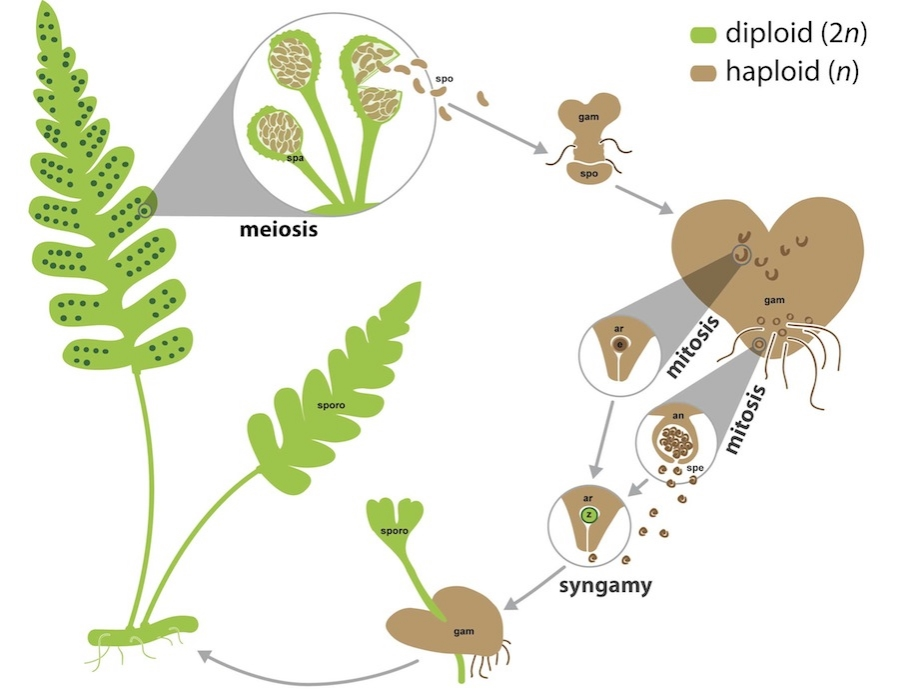
Simplified overview of the homosporous fern life cycle. Sporophyte tissues, which are often but not necessarily diploid, are shown in green. Gametophyte tissues and spores, which are often but not necessarily haploid, are shown in brown. Spores are generated by meiosis in sporangia. Gametes, both eggs and sperm, are generated by mitosis in archegonia and antheridia, respectively. For simplicity, fertilization is depicted between an egg and sperm from the same gametophyte, but fertilization is also likely to occur between gametes from different gametophytes that are derived from the same or different sporophytes. Abbreviations: an = antheridium; ar = archegonium; e = egg; gam = gametophyte; spa = sporangium; spe = sperm; spo = spore; sporo = sporophyte; z = zygote. Images are not to scale.
Ferns, like all land plants, have a life cycle that alternates between multicellular diploid (spore-producing sporophyte) and multicellular haploid (gamete-producing gametophyte) phases, punctuated by meiosis and fertilization. However, it is in the homosporous ferns that the manifestation of these two generations as independent entities is most extreme. Fern sporophytes are long-lived and complex branching plants composed of roots, stems, and leaves. Fern gametophytes, while smaller (often less than one centimeter) and having fewer tissue types, are mostly photosynthetic and can persist independently for multiple growing seasons, sometimes indefinitely. Thus, the fern sporophyte and gametophyte are two distinct organisms, varying in morphology, physiology, persistence, ecology, and (usually) chromosome number, but having a common genomic composition.

The phase-specific morphologies and functions of fern gametophytes and sporophytes result from different gene expression patterns, with some genes uniquely expressed in each phase. Working in collaboration with Dr. Joshua Der (California State University, Fullerton), we used RNA-Seq to generate transcript profiles of sporophyte and gametophyte tissue of the diploid fern species Polypodium amorphum (Sigel et al. 2018, Frontiers in Plant Science). Because of the unique biology of homosporous ferns, we were able to implement a biologically replicated, paired-sample experimental design whereby gametophytes are cultured from specific sporophyte individuals. Similar to the results of comparable studies in bryophytes and lycophytes, we found that nearly 90% overlap in the identity and expression levels of the genes expressed in both sporophytes and gametophytes, with less than 3% of genes uniquely expressed in either phase. Our findings suggest that shared patterns of phase-specific gene expression among seed-free plants likely reflect having relatively large, photosynthetic gametophytes, compared to the gametophytes of seed plants that are highly reduced. Phylogenetic analyses were used to further investigate the evolution of phase-specific expression for the phototropin, terpene synthase, and MADS-box gene families.